Powerful, superconducting magnet aids veterinary clinicians and students
A new piece of equipment at the Virginia-Maryland Regional College of Veterinary Medicine is letting clinicians and students take more detailed images of animals, both big and small.
The college’s Veterinary Teaching Hospital invested almost $700,000 on a magnetic resonance imaging (MRI) unit featuring a 1.5 Tesla magnet that is seven times more powerful than its predecessor. A Tesla is a measurement based on the strength of a magnetic field.
“Our new MRI unit not only lets us take higher quality images, but also increases their speed of acquisition,” said Dr. Dana Neelis, assistant professor of radiology in the Department of Small Animal Clinical Sciences. “Before we had the new magnet, we were limited to imaging small dogs and cats due to the size of the unit. Now we can accept any size of dog and can image certain areas on horses.”
The MRI unit uses a strong magnet to image hydrogen atoms within a patient’s body, creating slices of images of anatomic structures. MRI is most commonly used to diagnose abnormalities of the brain, spinal cord, and musculoskeletal system. It is also beneficial for surgical planning and oncologic purposes.
“The improved image quality with a stronger magnet helps us see small lesions that we may not have been able to appreciate with the weaker magnet,” Neelis said. “Each sequence of a scan now only takes one to five minutes, as opposed to the 10 to 15 minutes that it took before.”
A typical scan takes one hour for the brain and 1.5 hours for the spine. The Veterinary Teaching Hospital has already administered more than 200 scans, with an average of two or three scans a day.
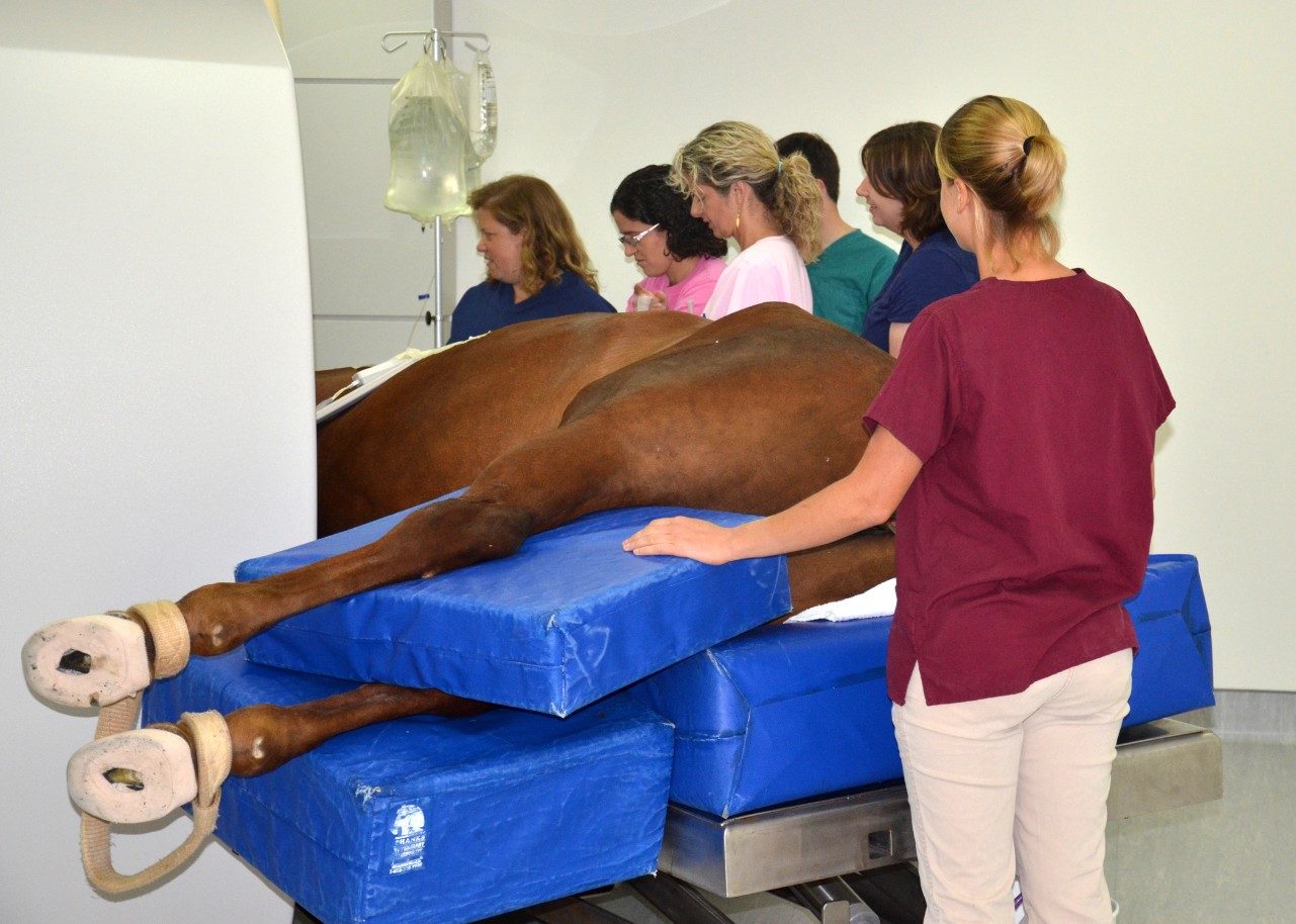
Dogs and cats are the most common patients to benefit from the college’s MRI unit, but it has seen a handful of horses. Like other four-legged patients, horses are put under general anesthesia before undergoing an MRI scan. Because of their large size, they need to be positioned on a specialized table and only certain areas — such as the feet, legs, and head — can fit into the magnet to be imaged.
According to Neelis, this MRI unit also has advancements in the types of sequences that can be performed. “We can now include 3-D sequences and diffusion-weighted imaging in our examinations, which were not available with the previous lower field magnet,” said Neelis, who explained that diffusion-weighted imaging is often used in human stroke patients to detect areas where blood supply to the brain may have been disturbed.
A room with copper shielding and specialized glass protects the superconducting magnet from stray radio signals that could interfere with its imaging capability. Liquid helium cools the magnet, maintaining the strong magnetic field, which stays on at all times. The renovation of the space to house the magnet cost more than $800,000. Additional equipment such as specialty monitors, anesthesia machines, MRI-compatible gurneys, and a stainless-steel table designed especially for equine MRI use brings the grand total for the MRI upgrade to more than $1.6 million.
This article was written by Michael Sutphin.

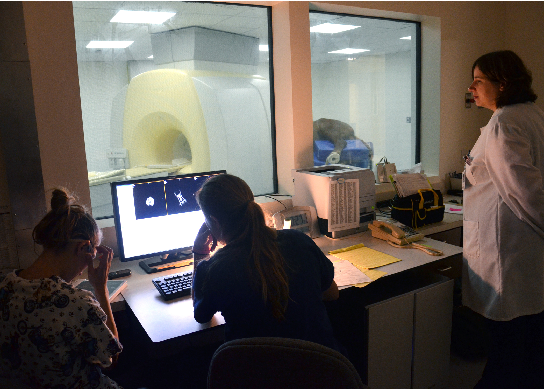
MRI control room
Hospital personnel monitor an MRI scan of a horse from a specialized control room.