


Seeing trees for the forest: Scientists find new aspects to visual system development
It’s not the destination that matters; it’s the journey – except when it comes to the brain.
Virginia Tech Carilion Research Institute scientists have found that the cells reaching from the eye to the brain form their branched endings differently, depending on where in the brain their endings terminate. The result is a single cell body with several terminals that look different.
In the retina, the cell body of a neuron extends an axon deep into the brain. This axon sends out branches searching for appropriate partner neurons. Once matched, they make a specialized intercellular connection called a synapse. Each branch ends in a different shape to accommodate its specific neuron partner.
The researchers used a rodent model for this analysis, but the results have possible implications for humans, said Michael Fox, the study’s lead scientist. The results were published recently in Neural Development.
“We’re interested in cells that live in the retina and how they project their axons into the brain,” said Fox, an associate professor at the Virginia Tech Carilion Research Institute. “In studying this we found that not all of the cell’s terminal endings appeared the same. And we think this is important as strategies to regenerate the injured nervous system progress.”
While regenerating retinal neurons and visual system circuits to restore vision in humans is still a long way off, this research is an important step, Fox said. The study is one of only a few that provide information about how different terminals from the same axon form, as well as how the synapse – where the opposed transmitting and receiving neurons exchange chemical signal information – forms.
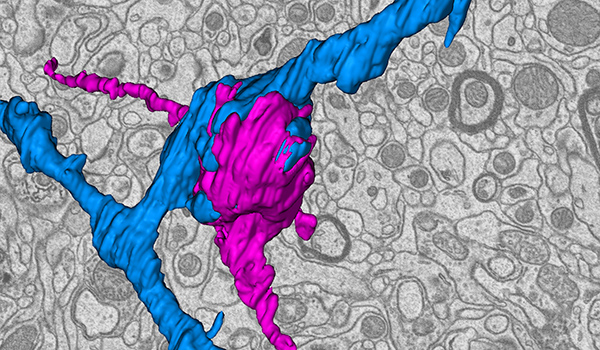
The researchers, including scientists from the University of Louisville in Kentucky, used an array of techniques to study the retinal terminals. They compared terminal endings in various areas of the brain and created 3-D reconstructions of the terminal structures from serial sections of high-resolution images taken with an electron microscope.
“Initially, we thought the terminals would all look and function similarly,” said Fox, who is also an associate professor of biological sciences in Virginia Tech’s College of Science. “The study showed us that a single axon could make very different terminals depending on which nuclei it’s targeting. This is important for the terminals’ function. Larger terminals are stronger.”
Not only did the scientists discover that the terminals changed shape and size depending on their locations, but they also found that the sizes of the terminals relative to one another changed with age. In young brains, the retinal terminals in different brain regions appeared equally sized, but as the synapses matured with age, those terminals developed unique morphologies and sizes.
With this information, Fox said, scientists can begin designing strategies to regenerate visual system circuits following neurotrauma or sight-related disorders, such as macular degeneration.
“These terminal details are especially important if you want to learn how to regenerate a specific terminal,” Fox added. “In thinking how to regenerate an injured visual system, it’s clear that retinal terminals in different parts of the brain will need to be tailored precisely.”
Written by Ashley WennersHerron.