


New, high-definition CT scanner arrives at Marion duPont Scott Equine Medical Center
New technology at Virginia Tech’s Marion duPont Scott Equine Medical Center (EMC) in Leesburg, Virginia, is offering enhanced imaging capabilities for equine patients.
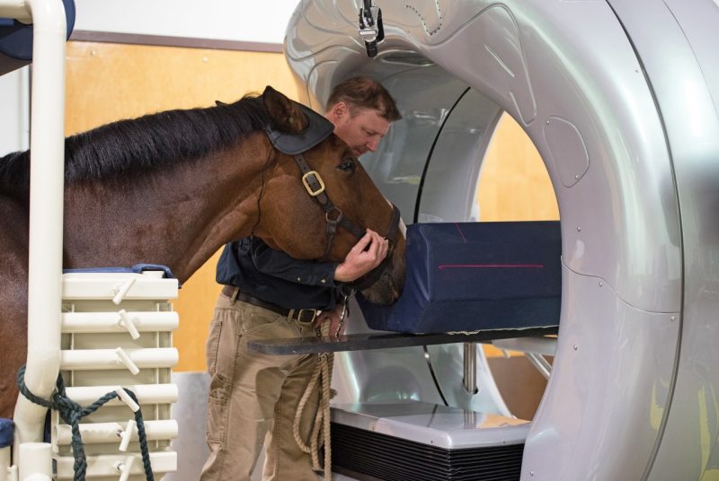
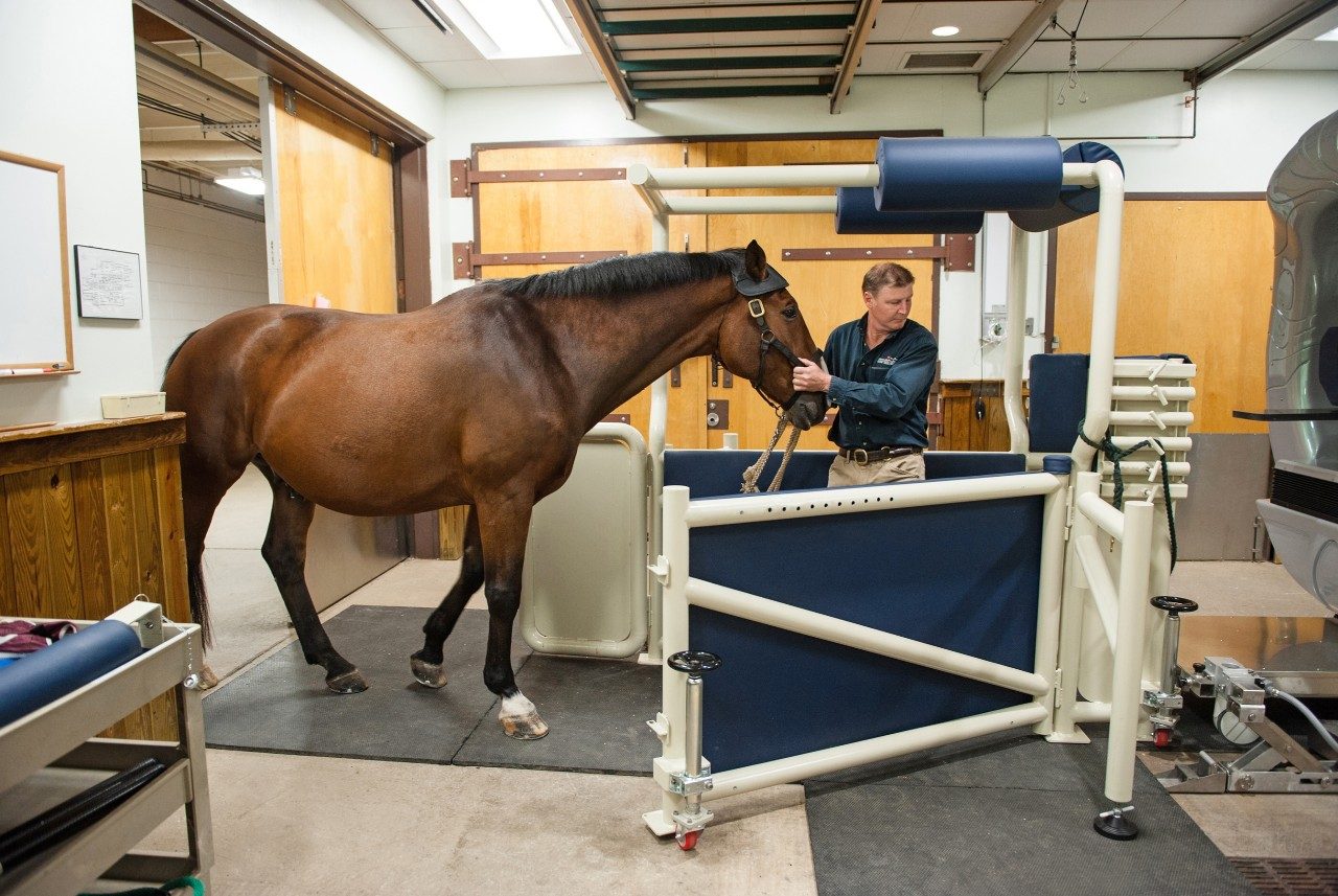
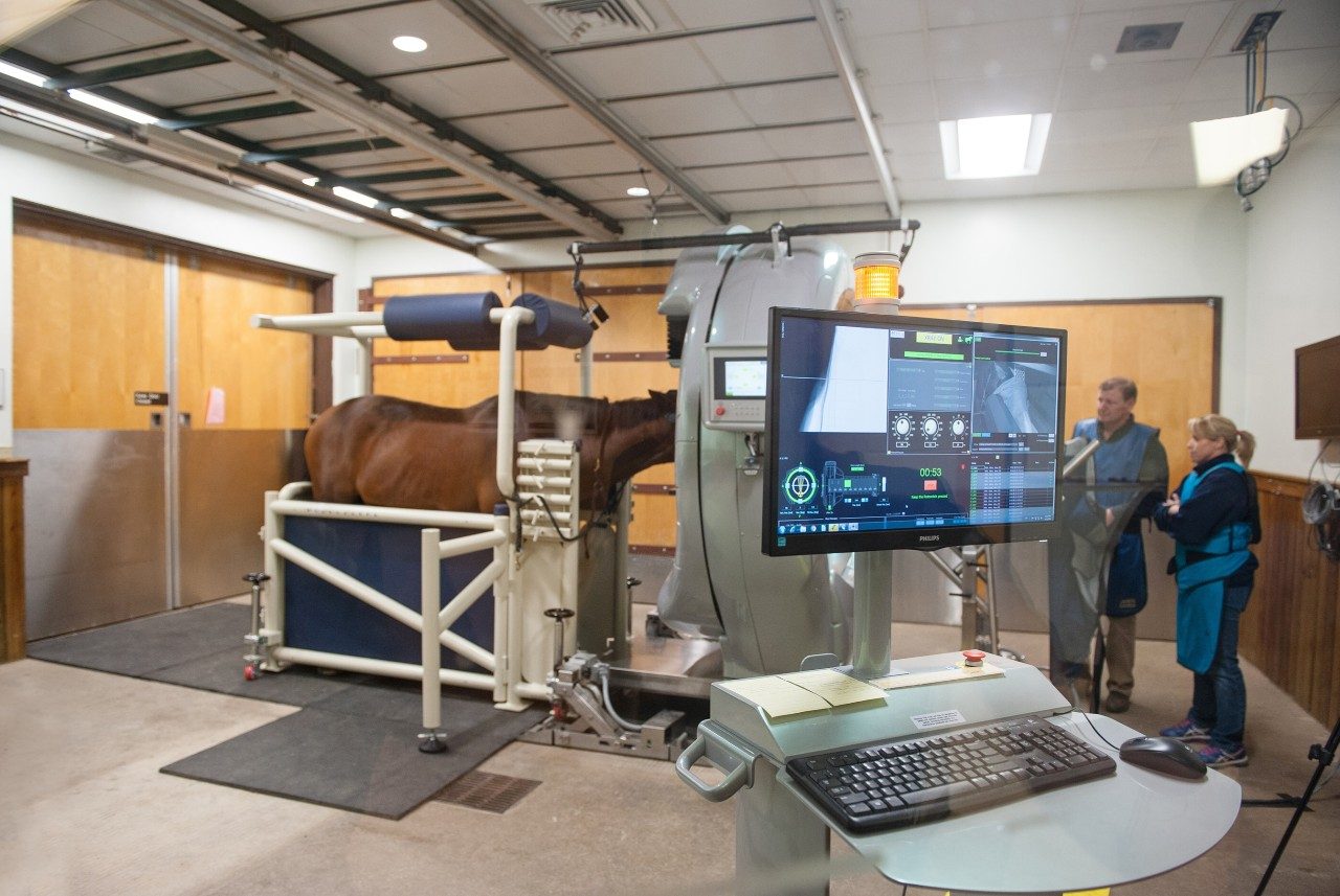
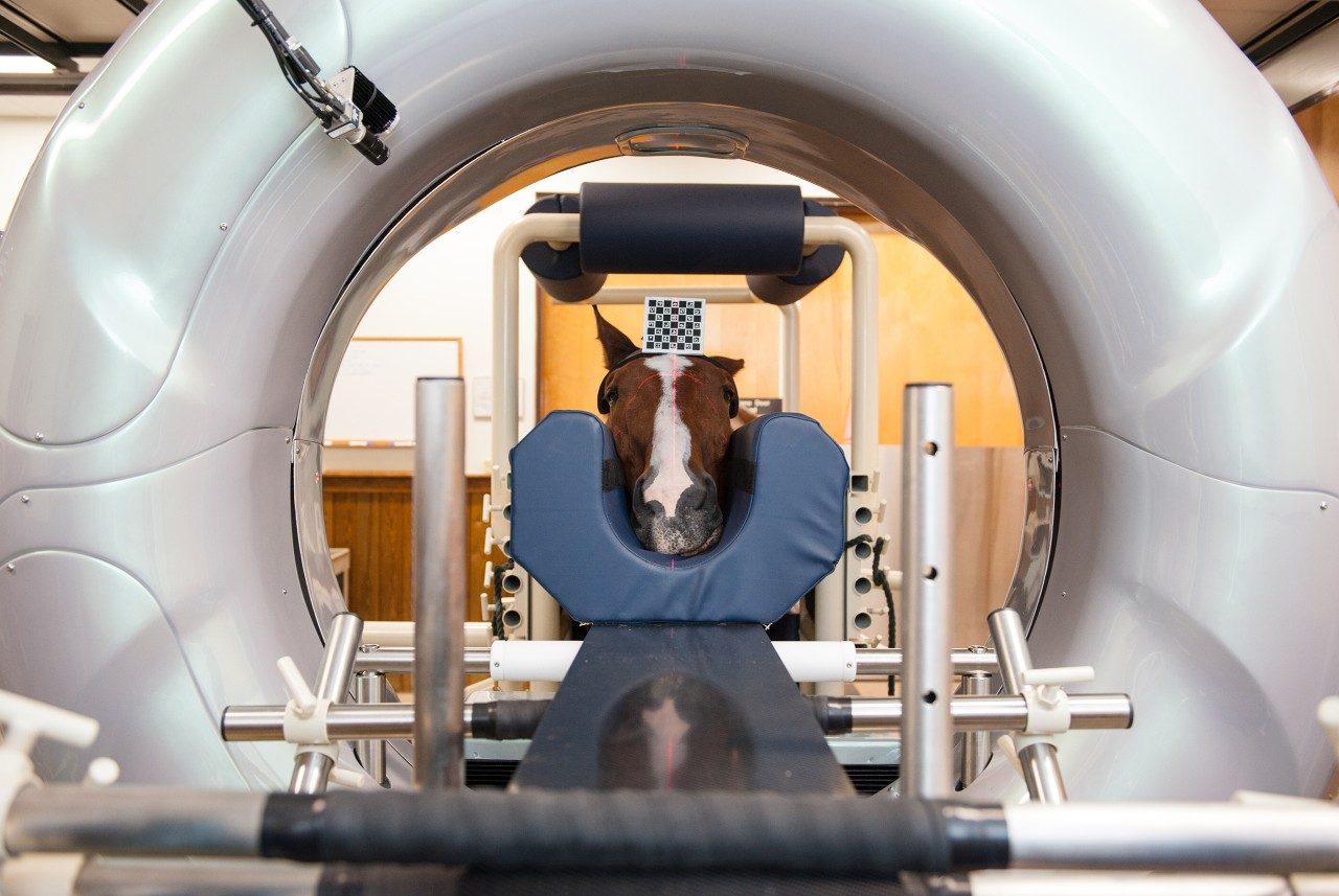
The Pegaso High-Definition CT, which is the first of its kind on the East Coast, allows veterinarians and staff at the center to perform high-definition CT scans on horses while standing or recumbent.
“We are very excited to offer this advanced technology for safer and clearer diagnoses of our equine patients,” said Mike Erskine, EMC director. The EMC is a part of the Virginia-Maryland College of Veterinary Medicine at Virginia Tech.
When a patient undergoes a CT (short for “computed tomography”) scan, multiple X-rays are taken at different angles to produce cross-sectional images, allowing a clinician to see inside the body without cutting. The new imaging technology at the EMC provides 3-D images at resolutions several orders of magnitude higher than a conventional CT but with vastly less radiation.
The purchase of the Pegaso scanner, which was created by Epica Medical Innovations, was made possible by a generous donation from the James Hale Steinman Foundation, as well as additional supporting gifts. There are only a handful of such scanners in use in the world — with others in Germany, Scotland, Colorado, and California.
The new technology enables CT scans to be performed on the head and neck of horses while standing and the distal limbs, stifle, and vertebrae to C7-T1 while recumbent.
“From my perspective, two aspects of this technology will be incredibly rewarding — the ability to image the stifle in three dimensions and the ability to image fractures, particularly complex fractures, in three dimensions for surgical repair,” said Jennifer Barrett, the Theodora Ayer Randolph Professor of Equine Surgery. “These additions greatly enhance our ability to provide the best treatment outcomes for orthopedic patients at the EMC.”
The CT scanner makes previously impossible-to-see areas clear, leading to highly detailed images, even with a horse moving during a study. It can also perform fluoroscopy, an imaging technique that creates real-time moving images.
“This imaging technology will be particularly beneficial in evaluating dental structures and paranasal sinuses, the neck, and orthopedic injuries involving the bones and joints,” Erskine said. “It is also well-suited to obtain detailed images of solid tumors.”
James Brown, clinical assistant professor of equine surgery, appreciates “the Pegaso’s ability to intricately display the anatomy of the horse’s head and neck in 3-D for clinical diagnostic purposes that cannot be accomplished with two-dimensional radiographs.”
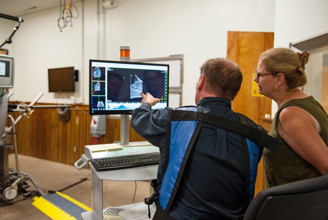
M. Norris Adams, clinical assistant professor in equine lameness and surgery, explained that the CT scanner can pinpoint surgical sites that would not be possible with any other imaging modality. “The ability to view 3-D images and subtract bone or soft tissue to concentrate on an area of interest with a single acquisition, which takes between 60 to 90 seconds to complete, has revolutionized our ability to diagnose and accurately identify disease and trauma sites,” Adams said.
Other imaging technologies available at the EMC include digital radiography, nuclear scintigraphy, digital ultrasound, and standing MRI. These diagnostic imaging services are popular with horse owners in the region.
“We currently have radiology and ultrasonography, including a standing MRI unit, that is quite advanced,” Erskine said, noting that clinicians perform approximately 170 MRI procedures every year. “We are expanding our imaging capabilities to help clinicians diagnose and treat lameness.”
The EMC is a full-service equine hospital located in Leesburg, Virginia, that offers advanced specialty care, 24-hour emergency treatment, and diagnostic services for all ages and breeds of horses. The veterinary college also treats horses at the Veterinary Teaching Hospital in Blacksburg and has an Equine Field Service there. In 2013, the teaching hospital in Blacksburg also invested in a powerful new MRI unit that can image certain parts of horses.
Read more about the Equine Medical Center in the latest issue of the college’s TRACKS magazine.


