New force measurement platform provides window to study cardiovascular disease
Cardiovascular disease is the number one cause of death in the United States. When the largest artery in the body, known as the aorta, is affected by disease, it can split or dilate, resulting in an aneurysm that can be fatal.
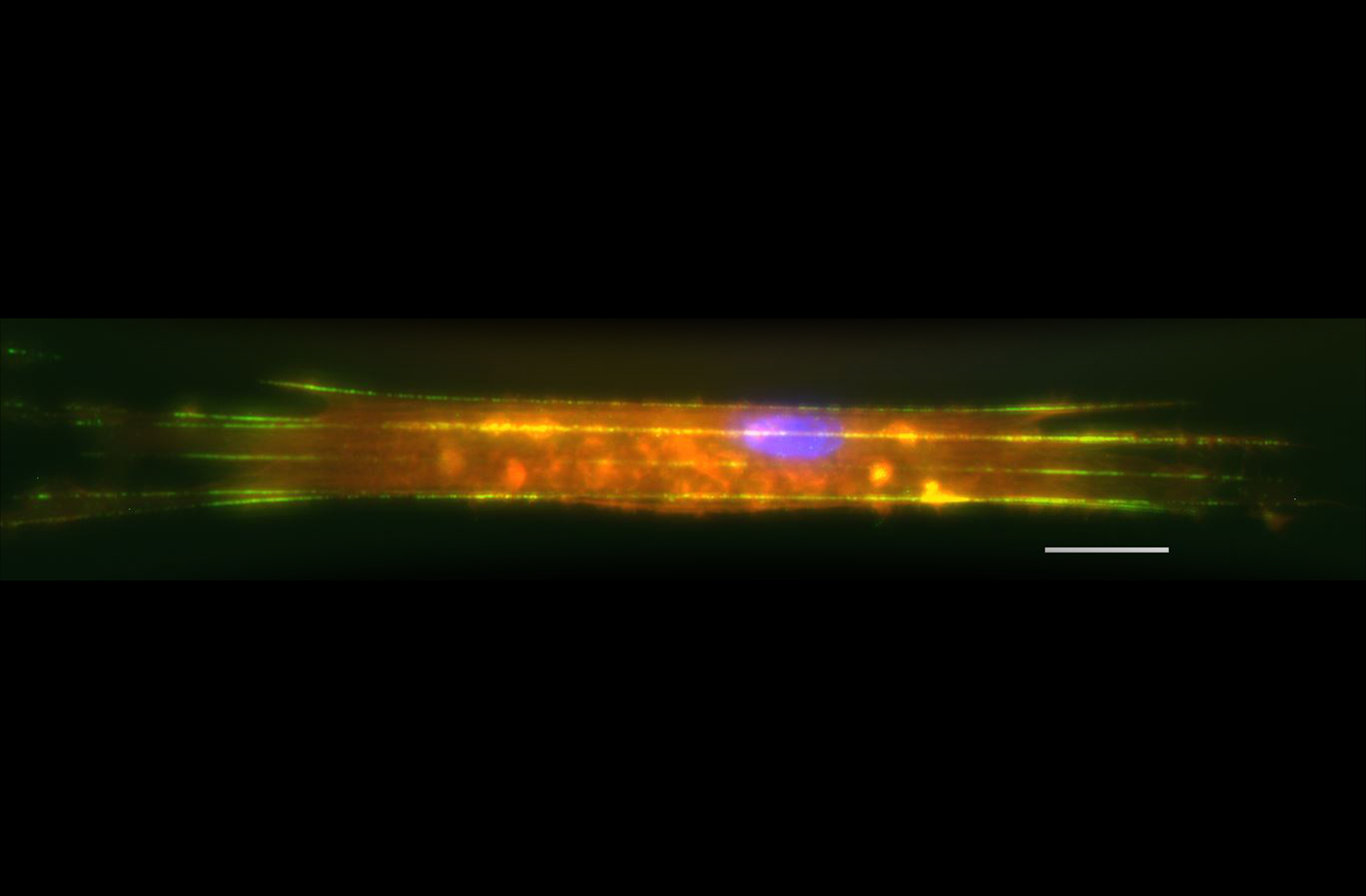
Virginia Tech and University of Pittsburgh School of Medicine researchers have developed a method to study the role of biomechanical forces and their disruption in diseased pathologies using relevant platforms that provide a window to study disease manifestation and progression. This platform, called nanonet force microscopy (NFM), is the first of its kind to measure single cell fiber forces, both under passive conditions and in the presence of disease conditions.
The findings have been published in “Forces” issue of the journal Molecular Biology of the Cell, in the article “Nanonet Force Microscopy for Measuring Forces in Single Smooth Muscle Cells of the Human Aorta."
Amrinder Nain, associate professor of mechanical engineering in the College of Engineering at Virginia Tech, pioneered NFM to utilize extracellular mimicking fibers in a controlled and repeatable manner. Together with Julie Phillippi, an assistant professor in the Department of Cardiothoracic Surgery at the University of Pittsburgh, they interrogate what the cells experience in the body by measuring individual cellular forces with a high level of precision.
Smooth muscle cells present in the walls of blood vessels undergo periodic expansion and contraction. The complex force signatures arising from this involve the interplay between the innate contractility of the cells and the forces exerted upon the cell by fibrous extracellular matrix, which structurally and functionally support these cells.
By altering the conditions of the matrix, such as fiber diameter, density, and spacing, or introducing an external force, NFM allowed them to understand how cells respond to the multitude of forces that they experience within tissues.
“Everything in nature exerts and experiences a physical force,” said Nain. “This platform measures both simultaneously.”
Phillippi studies the matrix and cell forces in blood vessel smooth muscle cells as a window to understanding aortic disease.
“The key idea behind our study is to show that disease mechanisms might be detectable at the single cell level,” Phillippi said.
Nain and Phillippi used cells from healthy individuals in the current study, but in the future, they plan to take advantage of the large repository of patient samples from healthy and diseased individuals established by Thomas Gleason, chief of the Division of Cardiac Surgery at the University of Pittsburgh, to determine force signatures for different types of cells in blood vessels under various conditions.
The technique has much broader applications as it represents a new method of disease modeling that could be built into drug testing platforms in the future. In a broader context, Nain thinks the ability to achieve precise control on fiber diameter, spacing, and orientation to mimic native fibrous environments, will allow NFM to interrogate the push and pulls in a cell’s journey in developmental, disease, and repair biology.
The expanded research team is composed of two undergraduate students, Christopher Delaughter of Mount Jackson, Virginia, and Matthew Apperson of Norfolk, Virginia; graduate student Alexander Hall, of Gastonia, North Carolina; and Kevin Sheets, former doctoral student, all of the Virginia Tech Department of Mechanical Engineering. The team also includes Patrick Chan, a clinical resident of the University of Pittsburgh.