Virginia Tech research could lead to better treatment of Duchenne muscular dystrophy
Mimicking human muscle function from dystrophic patients in a dystrophic mouse muscle in the lab could lead to enhanced treatment methods in patients.
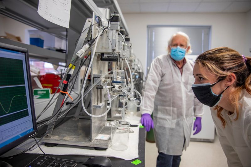
Kate Bukovec, right, analyzes data in the lab from research on Duchenne muscular dystrophy.
Duchenne muscular dystrophy, a rare form of muscular dystrophy that primarily affects boys, is uncurable. Although treatment that has revolved around symptom management has proved difficult, Virginia Tech researchers have found a way to potentially improve the available methods.
Duchenne muscular dystrophy is one of the most serious genetic diseases in children and more than 90 percent are in wheelchairs by age 11, with the average age of initial diagnosis at 5 years old. The ability to provide new, groundbreaking treatment methods based on mathematical models of dystrophic mouse muscles can test potential treatments for success rates with the potential to extend usable muscle function.
“Our approach provides a greater sensitivity for the assessment of disease progression, and our novel protocol could lead to better treatments for Duchenne muscular dystrophy,” said Robert Grange, an associate professor of human nutrition, foods, and exercise in the College of Agriculture and Life Sciences and affiliate faculty member in the Virginia Tech-Wake Forest School of Biomedical Engineering and Sciences and the Virginia Tech Center for Emerging, Zoonotic, and Arthropod-borne Pathogens.
The findings were published in a recent issue of the Journal of Applied Physiology, which is published by the American Physiological Society.
Human muscle function from dystrophic patients can be mimicked in a dystrophic mouse muscle in the lab. Muscle function during normal human walking, or gait, can be mathematically modeled, and these models can be used to mimic human muscle function in dystrophic mouse muscle. These models can test potential treatment methods through submersion in a bath with a special solution to keep the dystrophic mouse muscle alive that enables muscle contraction.
Another benefit of mimicking is that the models can be altered and applied to a variety of diseases that impact human gait.
“You can test gait in a mouse model of Alzheimer's or muscular dystrophy, so there aren't limits on this process,” said Kate Bukovec, the lead author on the paper and veterinary student in the Virginia-Maryland College of Veterinary Medicine. “I think what's even more important is that because we were able to run through this protocol with the soleus, a muscle in the lower calf, and we were also able to run through it with the extensor digitorum longus muscle in the leg when we get other simulation data, we can mimic different types of walking or running.”
Bukovec began working under Grange's mentorship during the veterinary college's Summer Veterinary Student Research Program in 2019. The program provides hands-on, mentor-guided biomedical and translational research experiences and builds opportunities for veterinary students across numerous career options.
The researchers, who are part of a larger team that includes researchers at the University of Virginia and engineers at Aurora Scientific, Inc., aim to address the broad scope of human movement in mice in the lab. Grange and Bukovec, his graduate assistant, studied the Duchenne muscular dystrophy aspect of the project.
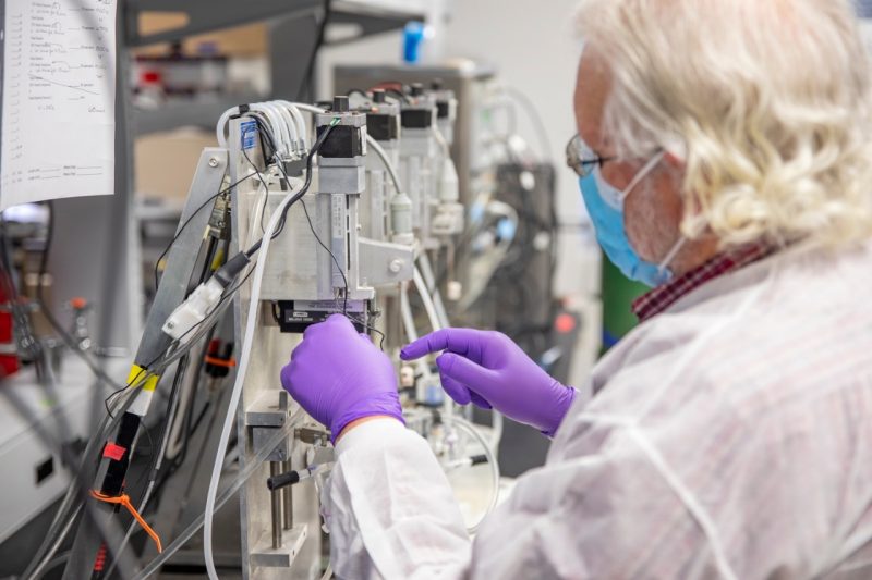
Grange prepares equipment from Aurora Scientific Inc. for use in the research on Duchenne muscular dystrophy.
A snapshot into the development process
Through their research, Grange and Bukovec were able to test human muscle length change and force production scaled to a mouse muscle.
The basic method the researchers typically use for assessing mouse muscle function has been around for decades: the electrodes in the special bath electrically stimulate contraction while the muscle length is changed. The unique part of the new method is that they could mimic – almost identically – the length changes that occurred in the human soleus muscle during gait in the mouse soleus muscle in the bath.
The next big issue was if the team could produce the forces that the muscle in the human soleus muscle produced during gait in the mouse muscle in the bath. The researchers collaborated with Aurora Scientific Inc. to modify the electrical stimulator to activate the mouse muscle in the bath to mimic almost exactly the force profiles of the human soleus. With the modification to the Aurora stimulator, both the appropriate force and length change patterns of the human soleus could be mimicked in the mouse soleus.
Application of the new method
Dynamic musculoskeletal simulation of walking was used to extract changes of force, length, and excitation of the human soleus during walking. To mimic human gait in much smaller mice, the changes in force and length were normalized and/or scaled based on muscle architectural parameters, and the changes in excitation – or electrical stimulation – were adjusted based on the actual force of each mouse soleus.
The processed changes in length and excitation were applied to the mouse soleus in the ex vivo protocol, and the normalized forces from the simulation were compared to ensure that comparable forces were produced in the ex vivo protocol. This protocol provides an assessment of simulated human movement in mouse muscle, including components of eccentric contractions.
“The protocol recapitulates both the patterns and magnitudes of physiological strains and stresses to provide greater insight into the effects of eccentric contractions on muscle function during gait,” Bukovec said. “Uniquely, the protocol incorporates a modified stimulator to deliver a physiological stimulation pattern that matches muscle force output during gait, which at present cannot be duplicated in existing acute eccentric protocols.”
The next steps
The research into mimicking dystrophic muscle function in mice is just a part of the overall goals of the research team writ large.
The long-term goal for the entire research team is a multiscale model, or a mathematical model, that starts with the micro aspects of the muscle in animals all way to the human scale. The models will build upon the research of Bukovec and Grange.
“This will let us determine in the lab why dystrophic muscles are more susceptible to injury when they are used to performing activities of daily living, such as when dystrophic boys walk downstairs and then test potential drug or gene therapy treatments to reduce or eliminate the injury,” said Grange, the director of the Virginia Tech Metabolism Core and a fellow in the Center for Transformative Research on Health Behaviors. “This mimicking approach could become a valuable method to assess in the lab if treatments are beneficial and should move on to the clinic to treat dystrophic boys, or if they are ineffective and should not be tried in the clinic on dystrophic boys.”
Ultimately, the team hopes to better understand how contractions during functional movements may translate to muscle damage and test the usability of potential treatments during the identical movements to limit damage to dystrophic muscles.
This work was supported by the National Institutes of Health. Facilities in the Virginia Tech Metabolism Core were used to conduct the studies.